Medical image analysis is the process of extracting meaningful information from medical images, often using computational methods. Some of the tasks for medical image analysis are visualization and exploration of 2D images and 3D volumes, segmentation, classification, registration, and 3D reconstruction of image data. The images for this analysis can be obtained from medical imaging modalities such as x-ray (2D and 3D), ultrasound, computed tomography (CT), magnetic resonance imaging (MRI), nuclear imaging (PET and SPECT), and microscopy. MATLAB®具有开发环境,并建立了用于建筑算法的分析和数据访问功能,用于医学图像分析。
DICOM file, which you can read, write, and anonymize in MATLAB.
可以使用医学图像分析来自动化或简化任务,例如计数和识别显微镜图像中的单元。例如,您可以分析和检测细胞中的癌症异常。对于重复或主观任务,计算医学图像分析可以消除由于人为错误而不一致。通过计算分析,您可以从坏死或测量血管中的氧饱和度分段肿瘤组织。
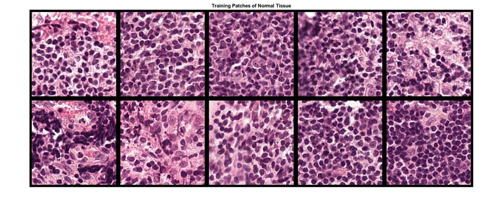
大型多分辨率图像的深度学习分类培训组织补丁。
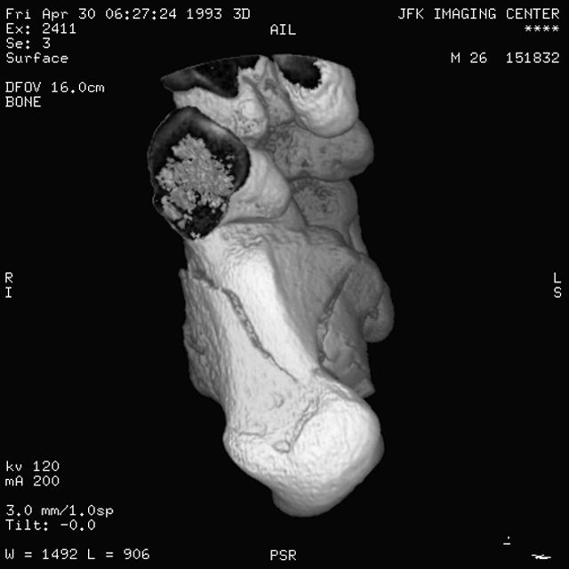
With medical image analysis, you can reconstruct a 3D representation from MRI images for calculating organ functions and other diagnostic measures
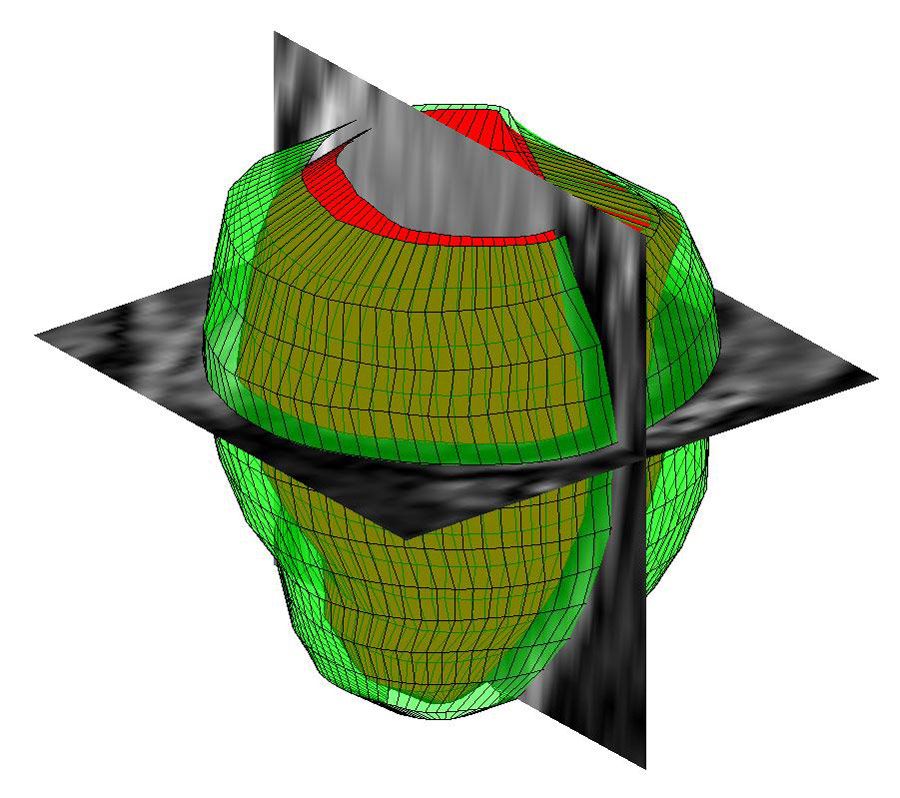
Three-dimensional geometrical reconstruction of the human left ventricle from MR images with MATLAB.
医学图像分析算法可以应用于大量数据,例如从可穿戴设备收集的数字健康数据。该算法可用于管理疾病和健康风险以及促进健康和福祉。
Matlab的医学图像分析
用matlab,你可以:
- 可视化和探索2D图像和3D卷
- Process very large multiresolution and high-resolution images
- Simplify medical image analysis tasks with built-in image segmentation algorithms
- Use deep learning techniques for classification
- 解析,加载,可视化和处理DICOM图像
In MATLAB, you can explore 3D volumetric data using the卷查看器应用程序。例如,您可以将人性大脑的MRI研究加载到批量查看器中,并探索显示大脑中发现的肿瘤的位置和类型的数据。
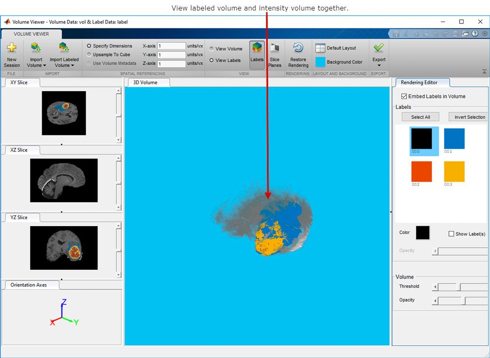
卷查看器应用程序,显示3D体积数据和3D标记的体积数据。
在数字病理学中,整个组织载玻片是成像和数字化。由此产生的整个幻灯片图像(WSIS)具有极高的分辨率。读取WSIS是一个挑战,因为图像无法加载到内存中,因此需要核心图像处理技术。马铃薯草极限对象可以存储和处理这种类型的大multiresolution image.
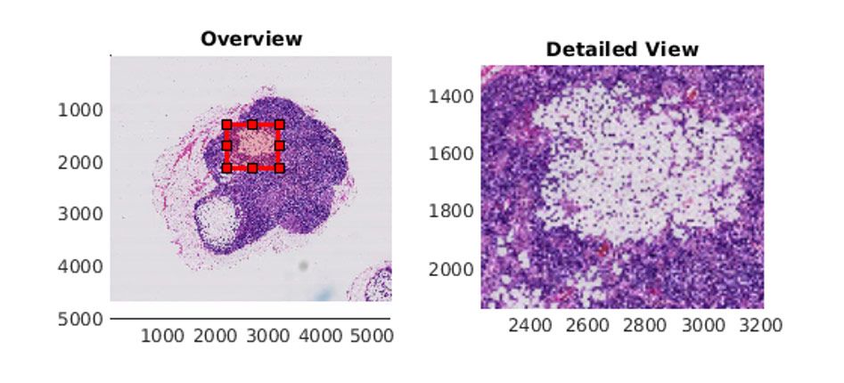
Image of a lymph node containing tumor tissue displayed withBigimageShow.in MATLAB.
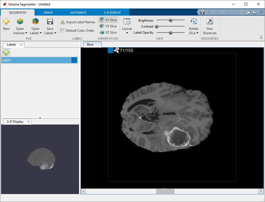
卷分段器应用程序,它显示卷的3D表示(在3D显示窗格中)和数据集的各个切片(在切片窗格中)。
通过MATLAB,您还可以使用深度学习方法来从3D医学图像执行脑肿瘤的语义分割。您可以设计和培训神经网络或使用掠夺网络。

使用MATLAB与标记的地面真理(左)和网络预测(右)进行脑组织中分段肿瘤。

