显微镜
MATLAB and Simulink for Microscope image analysis and instrument control
Scientists use MATLAB®从标准光学台面图像分析所有尺寸的显微镜图像到整个幻灯片分析。解剖学家,病理学家,微生物学家和生物医学专业人员使用MATLAB用于典型显微镜工作流程的所有步骤,包括预处理,细胞计数和分类,细胞跟踪,组织分割和疾病诊断。
MATLAB和图像处理工具箱™与计算机Vision Toolbox™,统计和机器学习工具箱™和深度学习工具箱™集成,使科学家能够在他们的研究中使用各种方法。在显微镜中也有一个强大的发展社区,并且该领域的专家正在创造更多的工具来帮助科学家与他们的特定任务。
“我们在Matlab中开发的算法并随着MathWorks顾问的帮助部署,使我们能够获得定量分析结果,避免人为错误,更有效地协作,可靠地再现成果,并且每年完成的可行性研究数量增加了两倍。”
Ryuta Saito, Mitsubishi Tanabe Pharma
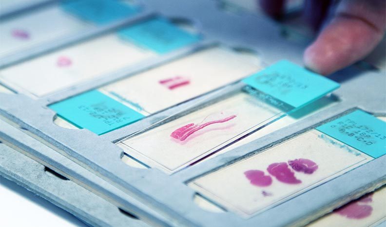
显微镜Image Analysis with MATLAB
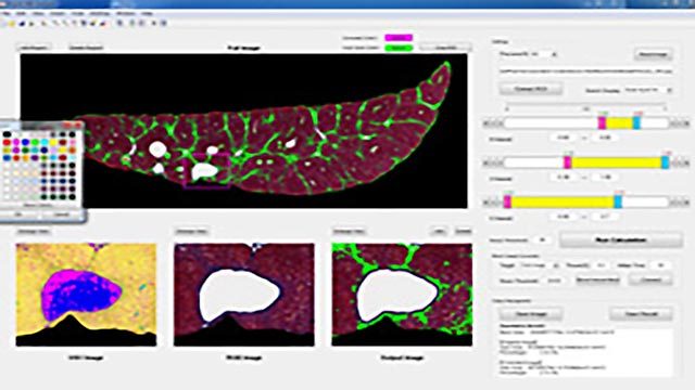
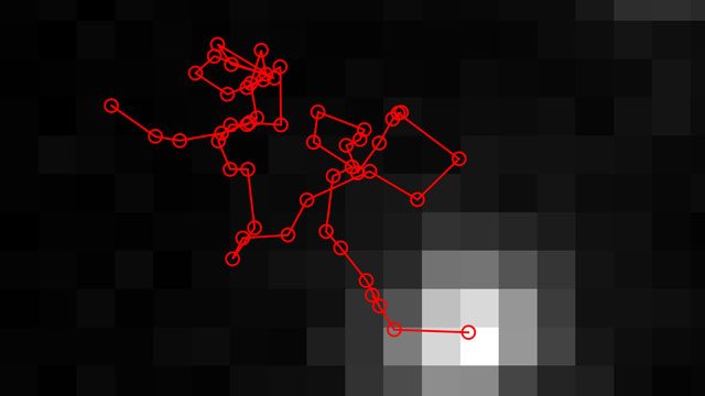
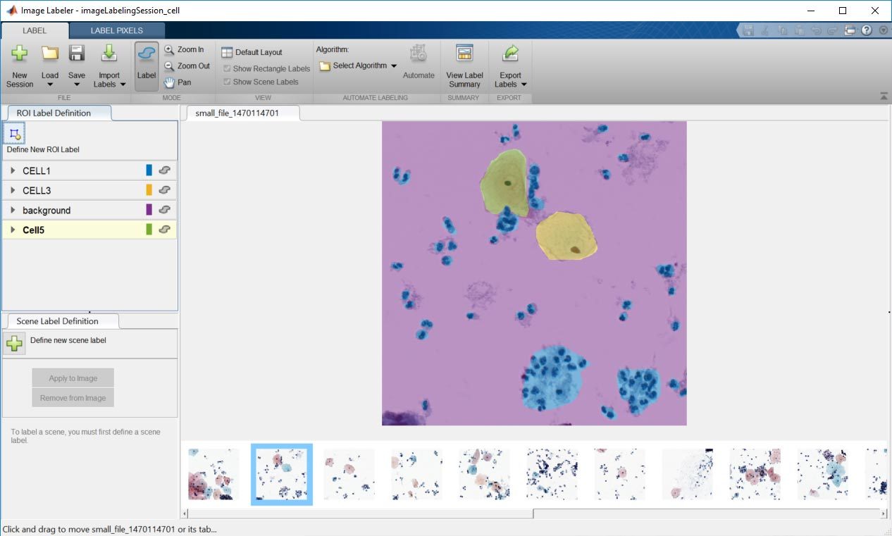
Scientists use MATLAB for quantitative analysis in microscopy workflows. Scientists can develop these workflows without writing code using apps like theImage Segmenter Appor theColor Thresholder App, and then automatically create documented code that replicates the interactive processing. Scientists also use morphological and general image processing to perform common microscopy tasks like cell segmentation, counting, and identification. There is a rich ecosystem of scientists developing tools for microscopy image analysis in MATLAB. For examples, see these文件交换中的显微镜工具。
Third-Party Toolboxes and Integration
Explore Products
Whole Slide Analysis with MATLAB
Bigimage, a new datatype for handling gigapixel whole slide images, was introduced to MATLAB in R2019b. Using this datatype, scientists can perform out-of-core operations on whole slide images using code developed for processing smaller microscopy images. This datatype integrates with Deep Learning Toolbox and enables high throughput whole slide analysis using deep learning. Scientists use MATLAB to predict outcomes, segment tissue, and analyze cancer in whole slide images.
Explore Products
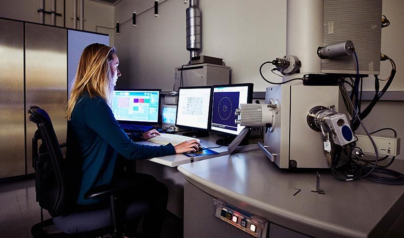
MATLAB显微镜仪器控制
Scientists and engineers can use MATLAB for control software on their microscopes as well as image acquisition, and general equipment control. Using a combination of all these tools, scientists can create fully functional microscopes that have high-level onboard image formation and analysis schemes. This can eliminate the need for a large data storage footprint and enable a complete workflow on one instrument.
Learn More
Explore Products

Get a Free Trial
30 days of exploration at your fingertips.

你是学生吗?
Get MATLAB and Simulink student software.